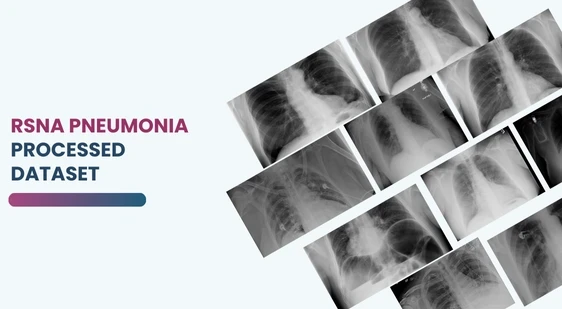
ML-Processed RSNA Pneumonia Dataset
ML-Processed RSNA Pneumonia Dataset
Datasets
ML-Processed RSNA Pneumonia Dataset
File
ML-Processed RSNA Pneumonia Dataset
Use Case
ML-Processed RSNA Pneumonia Dataset
Description
Discover the RSNA Pneumonia Detection Dataset, featuring pre-processed chest X-ray images, mask annotations, and detailed metadata for AI and machine learning in medical imaging

Description:
The RSNA Pneumonia Detection Dataset is a meticulously pre-processed version of the original RSNA Pneumonia Detection Challenge dataset, designed to simplify and accelerate machine learning workflows in healthcare. This dataset provides high-quality, pre-processed chest X-ray images (PNG format) with detailed annotations, making it ideal for developing AI models focused on pneumonia detection, classification, and medical imaging research.
Download Dataset
The RSNA Pneumonia Detection Dataset has been carefully pre-processed to streamline machine learning workflows, enabling faster and more efficient model training. Originally provided in DICOM format, the images have been converted into PNG format for compatibility and ease of use. The dataset preserves essential bounding box annotations by converting them into binary mask images, ensuring seamless integration into various training pipelines. Additionally, metadata, including patient details and bounding box coordinates, has been meticulously organized into CSV files, simplifying dataset exploration and feature engineering.
Folder Structure for Easy Navigation
The dataset is organized into a structured folder system, designed for straightforward usage:
1. Train
- Images: Pre-processed PNG images for training.
- Masks: Binary mask images corresponding to the bounding boxes in the training set.
2. Test
- Contains pre-processed PNG images designated for testing.
3. Metadata Files
- Train_metadata.csv: Includes patient IDs, bounding box coordinates, and pneumonia presence labels for the training set.
- Test_metadata.csv: Contains patient IDs and relevant details for the test set.
Key Data Fields
The dataset provides detailed annotations and metadata to support robust model development:
- patientId: Unique identifier for each patient or image.
- x, y, width, height: Bounding box dimensions specifying regions of interest.
- Target: Binary label indicating pneumonia presence (1 = Pneumonia, 0 = No Pneumonia).
- class: Categorization of conditions:
- Lung Opacity
- Normal
- No Lung Opacity / Not Normal
- age: Patient’s age at the time of imaging.
- sex: Patient’s gender (e.g., Male, Female).
- modality: Imaging method (e.g., X-ray, CT Scan).
- position: Patient’s position during imaging (e.g., AP for Anterior-Posterior, PA for Posterior-Anterior).
Dataset Distribution
The dataset includes a diverse range of cases, distributed as follows:
- No Lung Opacity / Not Normal: 11,821 images
- Normal: 8,851 images
- Lung Opacity: 9,555 images
This dataset is sourced from Kaggle.
Contact Us

Quality Data Creation

Guaranteed TAT

ISO 9001:2015, ISO/IEC 27001:2013 Certified

HIPAA Compliance

GDPR Compliance

Compliance and Security
Let's Discuss your Data collection Requirement With Us
To get a detailed estimation of requirements please reach us.
