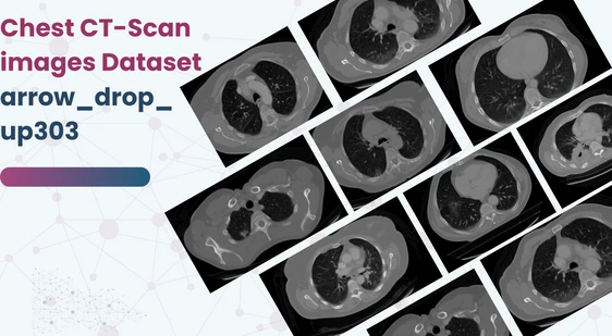
Chest CT-Scan images Dataset
Home » Dataset Download » Chest CT-Scan images Dataset
Chest CT-Scan images Dataset
Datasets
Chest CT-Scan images Dataset
File
Chest CT-Scan images Dataset
Use Case
Computer Vision
Description
It was a project about chest cancer detection using machine learning and deep leaning (CNN)

About Dataset
Data Story
This project is all about using artificial intelligence to detect chest cancer using a special kind of algorithm called Convolutional Neural Network (CNN). The AI model looks at chest X-ray images to figure out if someone has cancer or not.
Once the model makes a diagnosis, it also tells you what type of cancer it is and suggests the best way to treat it.
To make this AI model work, we needed a lot of data. I spent a long time searching for and gathering data from different sources. Then, I cleaned up the data to make sure it was all good for the CNN to use.
Now, with all the data ready, the AI can easily classify chest X-ray images and help doctors diagnose cancer more accurately.
Data
In this project, we’re using artificial intelligence to detect chest cancer from chest X-ray images. Instead of using a special format called “dcm,” the images are in common formats like JPG or PNG because that’s what the AI model can work with.
The data we collected includes images of three types of chest cancer: Adenocarcinoma, Large cell carcinoma, and Squamous cell carcinoma. There’s also a folder containing images of normal chest cells for comparison.
All these images are organized into different folders within a main folder called “Data.” Inside the “Data” folder, we have three subfolders: “test,” “train,” and “valid.” These folders help us separate the data for testing the AI model, training it, and validating its accuracy.
So, with this setup, we can train the AI to recognize different types of chest cancer and help doctors diagnose it more effectively.
test represent testing set
train represent training set
valid represent validation set
training set is 70%
testing set is 20%
validation set is 10%
Adenocarcinoma
Lung adenocarcinoma is the most prevalent form of lung cancer, constituting approximately 30% of all cases. Within non-small cell lung cancers (NSCLC), it’s even more predominant, accounting for about 40% of cases. Adenocarcinomas can also manifest in other parts of the body such as the breast, prostate, and colorectal areas. However, in the lungs, they primarily develop in the outer regions where glands produce mucus to aid in breathing. Symptoms of lung adenocarcinoma encompass coughing, hoarseness, weight loss, and weakness. If an individual experiences these symptoms, seeking medical attention for further evaluation is imperative.
Large cell carcinoma
Large-cell undifferentiated carcinoma is a swiftly growing type of lung cancer that spreads rapidly throughout the lung. It can develop in any part of the lung and is accountable for approximately 10 to 15 percent of all cases of non-small cell lung cancer (NSCLC). Detecting and treating this type of cancer early is crucial due to its rapid growth and spread.
Squamous cell carcinoma
Squamous cell lung cancer is usually found in the central part of the lung, where the larger airways connect to the trachea (windpipe) or in the main branches of the airways. It’s often associated with smoking and accounts for around 30% of all non-small cell lung cancers. In simpler terms, it’s a type of lung cancer that develops in specific areas of the lung and is more common in people who smoke. The last folder contains CT-Scan images of normal lungs, which are used for comparison when diagnosing lung cancer.
Acknowledgements
We wouldn’t be here without the help of others and the resources we found.
thanks for all of my team and the people who supported us
Inspiration
I want to hear all your feedback
This dataset is sourced from Kaggle.
Contact Us

Quality Data Creation

Guaranteed TAT

ISO 9001:2015, ISO/IEC 27001:2013 Certified

HIPAA Compliance

GDPR Compliance

Compliance and Security
Let's Discuss your Data collection Requirement With Us
To get a detailed estimation of requirements please reach us.
